1. Definition
What is an EEG Machine?
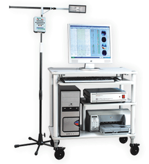
An Electroencephalography (EEG) machine is a sophisticated medical device designed to record and measure the electrical activity of the brain. It captures and amplifies the minute electrical impulses generated by neurons through electrodes placed on the scalp. The resulting record, called an electroencephalogram, provides crucial information about brain function and is fundamental in neurology, psychiatry, and sleep medicine.
How It Works
The working principle of an EEG machine is based on the detection of electrical potentials. Here’s how it works in simple terms:
- Signal Detection: When brain cells (neurons) communicate, they create tiny electrical charges. These charges create voltage fluctuations that can be detected on the scalp.
- Signal Amplification: These electrical signals are extremely weak (measured in microvolts – millionths of a volt). The EEG machine amplifies these signals millions of times.
- Signal Filtering: The amplified signals pass through filters that remove unwanted electrical interference (like muscle activity or power line interference).
- Signal Display and Recording: The processed signals are displayed as waveforms on a monitor and stored digitally for analysis.
The entire process is non-invasive and painless, similar to having your heart’s electrical activity measured by an ECG.
Key Components
- Electrodes: Small metal discs placed on the scalp to detect electrical activity. Modern systems typically use 19-256 electrodes.
- Amplifiers: Boost the weak brain signals to measurable levels while minimizing noise.
- Filters: Electronic circuits that remove unwanted frequencies (like muscle activity or AC power interference).
- Analog-to-Digital Converter (ADC): Converts the amplified analog signals into digital format for computer processing.
- Display Unit: Monitor showing real-time brainwave patterns.
- Recording System: Computer hardware and software for storing and analyzing EEG data.
- Impedance Checker: Measures electrode-skin contact quality to ensure accurate recordings.
- Headboxes/Electrode Caps: Interface between electrodes and amplifiers, often organized in caps for easy placement.
- Stimulators: Devices that deliver visual, auditory, or sensory stimuli during specialized EEG tests.
2. Uses
Clinical Applications
EEG machines serve multiple critical functions in clinical medicine:
- Epilepsy Diagnosis and Management: The primary use, helping identify seizure types, locate seizure foci, and monitor treatment effectiveness.
- Sleep Disorders: Used in polysomnography to study sleep stages and diagnose disorders like narcolepsy and sleep apnea.
- Brain Injury Assessment: Evaluates brain function after trauma, stroke, or hypoxia.
- Encephalopathy Evaluation: Assesses brain dysfunction from metabolic disturbances, infections, or toxins.
- Coma and Brain Death Determination: Helps evaluate consciousness levels and confirm brain death in some protocols.
- Dementia and Neurodegenerative Disorders: Assists in differentiating types of dementia.
- Surgical Planning: Used in epilepsy surgery to identify areas to be removed while preserving essential functions.
- ICU Monitoring: Continuous EEG (cEEG) monitors for non-convulsive seizures in critically ill patients.
- Research Applications: Studies brain development, cognitive processes, and psychiatric disorders.
Who Uses It
- Neurologists (particularly epileptologists)
- Neurophysiologists
- Psychiatrists
- Sleep Specialists
- EEG Technologists/Technicians
- Neurosurgeons
- Critical Care Physicians
- Research Scientists
Departments/Settings
- Neurology Departments
- Epilepsy Monitoring Units
- Sleep Laboratories
- Intensive Care Units (ICUs)
- Emergency Departments
- Operating Rooms (for intraoperative monitoring)
- Psychiatry Departments
- Rehabilitation Centers
- Academic and Research Institutions
- Ambulatory/Home EEG Settings
3. Technical Specifications
Typical Specifications
- Channels: 19-256 recording channels (standard clinical EEG uses 21-32 channels)
- Sampling Rate: 200-5000 Hz per channel
- Input Impedance: >100 MΩ
- Common Mode Rejection Ratio (CMRR): >100 dB
- Noise Level: <0.5 µV peak-to-peak
- Bandwidth: 0.1-100 Hz (typical), up to 500 Hz for high-frequency oscillations
- Resolution: 16-24 bits
- Display: High-resolution monitors, often with touchscreen capability
- Connectivity: Ethernet, Wi-Fi, Bluetooth, USB ports
- Battery Backup: 2-8 hours for portable units
Variants & Sizes
- Routine/Diagnostic EEG Machines: Standard systems for short recordings (20-60 minutes)
- Ambulatory EEG Systems: Portable devices for 24-72 hour home monitoring
- Video-EEG Monitoring Systems: Combined EEG and video recording for epilepsy monitoring
- ICU/cEEG Systems: For continuous monitoring in critical care
- Portable/Handheld EEG: Compact devices for emergency or bedside use
- High-Density EEG: Systems with 64-256 channels for precise source localization
- Neonatal EEG: Specialized systems for newborns and infants
Materials & Features
Construction Materials:
- Medical-grade plastics and metals
- Silver/silver chloride or gold electrodes
- Shielded cables to reduce interference
- Antimicrobial surfaces for infection control
Advanced Features:
- Automatic Impedance Checking: Real-time electrode contact quality monitoring
- Digital Video Synchronization: Precise alignment of EEG and video data
- Automatic Event Detection: Algorithms to identify seizures, spikes, and other patterns
- Source Localization Software: 3D brain mapping capabilities
- Quantitative EEG (qEEG): Advanced analytical tools
- Network Connectivity: For remote monitoring and telemedicine
- Mobile Integration: Tablet/smartphone control and viewing
- Cloud Storage and Analysis: Secure data storage and processing
Notable Models
- Nicolet EEG Systems (Natus Medical)
- Neurofax EEG-1200 (Nihon Kohden)
- BrainAmp Series (Brain Products)
- g.USBamp and g.HIAMP (g.tec medical engineering)
- Mizar EEG Systems (EBNeuro)
- NeuroScan Series (Compumedics)
- Xltek EEG Systems (Natus Medical)
- Micromed Systems (Micromed S.p.A.)
- Walter Graphtek Systems (Walter Graphtek GmbH)
- Cadwell EEG Systems (Cadwell Industries)
4. Benefits & Risks
Advantages
- Non-invasive and Painless: No needles or incisions required
- High Temporal Resolution: Captures changes in milliseconds
- Relatively Low Cost: Compared to other neuroimaging like MRI or PET
- Portable Options Available: Can be used at bedside or in patient’s home
- Real-time Monitoring: Immediate feedback on brain activity
- Safe for Repeated Use: No radiation exposure
- Excellent for Seizure Detection: Gold standard for epilepsy diagnosis
- Versatile Applications: From clinical to research settings
- Established Standards: Well-understood patterns and interpretations
Limitations
- Poor Spatial Resolution: Cannot precisely locate deep brain activity
- Signal Attenuation: Skull and scalp distort and weaken signals
- Susceptible to Artifacts: Affected by muscle movement, eye blinks, electrical interference
- Requires Technical Expertise: Proper electrode placement and interpretation need training
- Limited Functional Information: Shows electrical activity but not brain structure or metabolism
- Patient Cooperation Needed: Movement artifacts can ruin recordings
- Time-Consuming Setup: Proper electrode placement takes 20-45 minutes
Safety Concerns & Warnings
- Electrical Safety: Must comply with IEC 60601 standards to prevent electrical shock
- Infection Control: Proper electrode cleaning between patients
- Fall Risk: Patients connected to EEG may have limited mobility
- Seizure Risk During Testing: Especially during activation procedures
- Skin Irritation: From electrode paste or adhesives
- Allergic Reactions: To electrode materials or conductive paste
- Data Security: Protected health information must be secured
- Equipment Interference: With other medical devices (rare but possible)
Contraindications
- Open Wounds or Infections on the scalp where electrodes need placement
- Severe Scalp Conditions like psoriasis or eczema that prevent electrode adhesion
- Uncooperative Patients who cannot remain still (unless sedated)
- Emergency Situations where the EEG setup would delay critical care (relative contraindication)
- Known Severe Allergies to electrode materials or conductive media
5. Regulation
FDA Class
Class II Medical Device (special controls required)
- Regulation Number: 21 CFR 882.1400
- Product Code: GWM (Electroencephalograph)
- Requires 510(k) premarket notification in most cases
- Subject to performance standards, postmarket surveillance, and special labeling
EU MDR Class
Class IIa Medical Device under EU MDR 2017/745
- Rule 10: Active therapeutic devices and devices for diagnosis/monitoring
- Requires Notified Body assessment
- Must meet general safety and performance requirements (Annex I)
- Requires CE marking with Notified Body number
CDSCO Category
Category B (Moderate risk) under Medical Device Rules, 2017 (India)
- Registration required with Central Drugs Standard Control Organization
- Subject to conformity assessment by Notified Body
- Must comply with ISO standards and CDSCO labeling requirements
PMDA Notes
- Regulated as Class II Medical Devices under Pharmaceutical and Medical Device Act (Japan)
- Requires Marketing Authorization from PMDA
- Must comply with Japanese Industrial Standards (JIS) and Pharmaceutical Affairs Law
- Often requires clinical data from Japanese population
ISO/IEC Standards
- ISO 80601-2-26: Particular requirements for basic safety and essential performance of EEG equipment
- IEC 60601-1: General requirements for basic safety and essential performance
- IEC 60601-1-2: Electromagnetic compatibility requirements
- ISO 13485: Quality management systems for medical devices
- IEC 62304: Medical device software lifecycle processes
- ISO 14971: Application of risk management to medical devices
- IEC 62366: Usability engineering for medical devices
- ISO 10993: Biological evaluation of medical devices
6. Maintenance
Cleaning & Sterilization
Daily/After Each Use:
- Wipe external surfaces with hospital-grade disinfectant (70% isopropyl alcohol or approved disinfectant wipes)
- Clean electrode surfaces with mild soap solution or specialized electrode cleaner
- Rinse with distilled water and dry thoroughly
- For disposable electrodes: discard after single use according to biomedical waste protocols
Weekly/Monthly:
- Deep clean cable connectors with appropriate cleaning solutions
- Check cables for fraying or damage
- Clean ventilation fans and filters
Sterilization Note: Most EEG components are not suitable for autoclaving. Only use manufacturer-approved cleaning agents.
Reprocessing
- Electrodes: Disposable electrodes are single-use only. Reusable electrodes require thorough cleaning and disinfection between patients.
- Electrode Caps: Clean with mild detergent, disinfect with approved solution, rinse thoroughly, air dry.
- Cables and Headboxes: Wipe with disinfectant; avoid liquid ingress into connectors.
Calibration
- Daily/Before Each Use: Biological calibration using “calibration signal” feature
- Monthly: Check amplifier gain, filter settings, and impedance measurement accuracy
- Quarterly: Full technical calibration by trained technician
- Annual: Comprehensive calibration by manufacturer or certified service provider
- After Major Repairs: Always recalibrate
Calibration should follow manufacturer specifications and be documented in equipment log.
Storage
- Environment: Store in clean, dry area with temperature 15-30°C and humidity 30-70%
- Cables: Store loosely coiled, avoid sharp bends
- Electrodes: Keep in sealed containers to prevent oxidation
- Batteries: Maintain charge at 40-60% for long-term storage; cycle every 3 months
- Protection: Use dust covers when not in use
- Transport: Use original packaging or padded cases for portable units
7. Procurement Guide
How to Select the Device
Consider these factors:
- Clinical Needs: Epilepsy monitoring, sleep studies, ICU monitoring, or general neurology?
- Patient Volume: Number of beds/patients to be served
- Technician Expertise: Complexity of operation and analysis required
- Space Availability: Fixed installation vs. portable/mobile needs
- Budget Constraints: Initial cost vs. total cost of ownership
- Future Expansion: Scalability and upgrade options
- Integration Requirements: Compatibility with existing hospital systems
Quality Factors
- Signal Quality: High CMRR, low noise, appropriate sampling rate
- Reliability: Mean time between failures, warranty terms
- Ease of Use: Intuitive interface, setup time, workflow integration
- Service Support: Manufacturer support availability, response time
- Software Capabilities: Analysis tools, reporting features, updates
- Build Quality: Durability, materials, design
- Patient Comfort: Electrode systems, recording comfort
Certifications to Look For
- CE Marking (for European markets)
- FDA 510(k) Clearance or Premarket Approval (for US market)
- ISO 13485 Certification (quality management)
- IEC 60601 Series Compliance (electrical safety)
- Regional Certifications (PMDA for Japan, CDSCO for India, etc.)
- Cybersecurity Certifications (for network-connected devices)
Compatibility Considerations
- Hospital Information Systems: HL7 interface for EMR integration
- PACS Compatibility: DICOM support for image storage
- Network Infrastructure: Ethernet/Wi-Fi compatibility
- Other Neurodiagnostic Equipment: Compatibility with EMG, evoked potentials, etc.
- Power Systems: Voltage requirements, battery backup
- Accessories: Electrodes, pastes, caps from multiple suppliers
Typical Pricing Range
- Basic Diagnostic EEG: $15,000 – $40,000
- Video-EEG Monitoring Systems: $50,000 – $150,000
- Ambulatory EEG Systems: $20,000 – $60,000
- High-Density Research Systems: $80,000 – $300,000
- Portable/Handheld EEG: $5,000 – $25,000
Prices vary significantly based on channels, features, and manufacturer.
8. Top 10 Manufacturers (Worldwide)
- Natus Medical Incorporated (USA)
- Headquarters: Pleasanton, California, USA
- Profile: Global leader in neurodiagnostics, formed through mergers of Nicolet, XLTEK, and others
- Notable Lines: Nicolet EEG, Xltek EEG, Olympic EEG
- Nihon Kohden Corporation (Japan)
- Headquarters: Tokyo, Japan
- Profile: Major Japanese manufacturer with strong global presence in patient monitoring and neurodiagnostics
- Notable Lines: Neurofax EEG-1200, EEG-2200
- Compumedics Limited (Australia)
- Headquarters: Melbourne, Australia
- Profile: Specialized in sleep diagnostics and neurodiagnostics with strong R&D focus
- Notable Lines: Grael, NeuroScan, SonmoStar
- Brain Products GmbH (Germany)
- Headquarters: Gilching, Germany
- Profile: High-end research-focused EEG systems, known for excellent signal quality
- Notable Lines: BrainAmp, actiCHamp, LiveAmp
- Cadwell Industries, Inc. (USA)
- Headquarters: Kennewick, Washington, USA
- Profile: Family-owned company specializing in neurodiagnostic equipment
- Notable Lines: Easy II, Easy III EEG
- Medtronic plc (Ireland)
- Headquarters: Dublin, Ireland (operational in Minnesota, USA)
- Profile: Medical technology giant with EEG through acquisitions
- Notable Lines: EEG products through Covidien integration
- Micromed S.p.A. (Italy)
- Headquarters: Mogliano Veneto, Italy
- Profile: Italian manufacturer with strong European presence
- Notable Lines: Micromed EEG Systems, SAM 32
- EBNeuro S.p.A. (Italy)
- Headquarters: Florence, Italy
- Profile: Specialized in clinical neurophysiology equipment
- Notable Lines: Mizar, Galileo EEG Systems
- g.tec medical engineering GmbH (Austria)
- Headquarters: Schiedlberg, Austria
- Profile: Focus on research and brain-computer interface applications
- Notable Lines: g.USBamp, g.HIAMP
- ANT Neuro B.V. (Netherlands)
- Headquarters: Hengelo, Netherlands
- Profile: Specialized in EEG solutions for research and clinical applications
- Notable Lines: eego series, WaveGuard caps
9. Top 10 Exporting Countries (Latest Year)
Based on 2023 export data for EEG machines (HS Code 901819):
- United States
- Export Value: $320 million
- Notes: Leading exporter with major manufacturers like Natus and Cadwell
- Germany
- Export Value: $185 million
- Notes: Strong engineering base, home to Brain Products and other specialized manufacturers
- Japan
- Export Value: $150 million
- Notes: Nihon Kohden dominates Asian markets with high-quality systems
- Netherlands
- Export Value: $95 million
- Notes: Major distribution hub for European market with growing domestic manufacturers
- China
- Export Value: $85 million
- Notes: Rapidly growing exporter with improving technology and competitive pricing
- Italy
- Export Value: $70 million
- Notes: Home to several specialized manufacturers like Micromed and EBNeuro
- United Kingdom
- Export Value: $55 million
- Notes: Strong research sector driving specialized EEG exports
- South Korea
- Export Value: $45 million
- Notes: Growing presence with advanced digital technology integration
- France
- Export Value: $40 million
- Notes: Traditional strength in medical technology with specialized EEG applications
- Switzerland
- Export Value: $35 million
- Notes: High-precision manufacturing for specialized clinical and research applications
10. Market Trends
Current Global Trends
- Increasing Epilepsy Prevalence: Rising neurological disorders driving market growth
- Aging Population: Higher incidence of dementia, stroke, and age-related neurological conditions
- Portability Demand: Shift toward ambulatory and home-based EEG monitoring
- Tele-neurology Expansion: Remote EEG reading and telemedicine integration
- Emerging Markets Growth: Increasing healthcare expenditure in Asia, Latin America, and Africa
- Consolidation: Larger companies acquiring specialized EEG manufacturers
- Value-Based Healthcare: Focus on cost-effective diagnosis and monitoring
New Technologies
- Wireless EEG Systems: Bluetooth and Wi-Fi enabled devices for improved patient mobility
- Dry Electrodes: Eliminating conductive gel for easier setup
- Artificial Intelligence: Machine learning algorithms for automatic pattern recognition and diagnosis
- High-Density EEG: 256+ channels for improved spatial resolution
- Wearable EEG: Discreet, long-term monitoring devices
- Quantitative EEG (qEEG): Advanced analytical techniques for better interpretation
- Source Localization Advances: Improved algorithms for precise brain mapping
- Cloud-Based Platforms: Secure data storage and collaborative analysis tools
Demand Drivers
- Rising Neurological Disorder Prevalence: Epilepsy affects 50 million people worldwide (WHO)
- Technological Advancements: Improved accuracy and ease of use
- Growing Awareness: Better diagnosis rates for neurological conditions
- Healthcare Infrastructure Development: Especially in emerging economies
- Research Funding Increase: Neuroscience research driving equipment needs
- Home Healthcare Trend: Preference for home-based diagnostics
- Regulatory Support: Clearer pathways for device approval and reimbursement
Future Insights
- Integration with Other Modalities: Combined EEG-fMRI, EEG-MEG systems
- Real-time Closed-loop Systems: For seizure prediction and intervention
- Consumer EEG Devices: For wellness and cognitive enhancement monitoring
- Advanced Biomarkers: For early diagnosis of neurodegenerative diseases
- Personalized Medicine: EEG-guided treatment planning
- Global Standardization: Harmonized protocols and interpretation guidelines
- Sustainability Focus: Eco-friendly materials and energy-efficient designs
11. Training
Required Competency
For EEG Technologists:
- Formal education in electroneurodiagnostics or related field
- Certification (R. EEG T. in US, equivalent elsewhere)
- Understanding of neuroanatomy and neurophysiology
- Proficiency in the 10-20 electrode placement system
- Recognition of normal and abnormal EEG patterns
- Knowledge of activation procedures and safety protocols
- Basic troubleshooting skills
For Physicians/Interpreters:
- Neurology or clinical neurophysiology specialization
- Fellowship training in EEG interpretation
- Board certification in clinical neurophysiology (preferred)
- Continuing education in evolving EEG technologies
Common User Errors
- Poor Electrode Application: Incorrect placement or high impedance
- Inadequate Patient Preparation: Failure to explain procedure, leading to movement artifacts
- Incorrect Montage Selection: Using inappropriate montages for clinical question
- Filter Misuse: Over-filtering or under-filtering signals
- Improper Calibration: Skipping daily calibration checks
- Artifact Misinterpretation: Mistaking muscle or eye movement for cerebral activity
- Inadequate Documentation: Poor recording of patient state, medications, or clinical events
- Safety Oversights: Failure to monitor patients during activation procedures
Best-Practice Tips
- Patient Preparation: Explain procedure thoroughly, ensure comfort, minimize hair products
- Systematic Approach: Follow standardized setup protocols every time
- Continuous Monitoring: Watch for patient state changes and document accordingly
- Quality Checks: Verify impedance, calibration, and signal quality throughout recording
- Comprehensive Documentation: Record medications, patient state, and clinical events
- Regular Maintenance: Follow manufacturer guidelines for cleaning and calibration
- Continuing Education: Stay updated on new techniques and guidelines
- Team Communication: Collaborate with referring physicians for optimal study design
12. Frequently Asked Questions (FAQs)
Q1: Is an EEG test painful or dangerous?
A: No, EEG is completely painless and non-invasive. It records your brain’s natural electrical activity without sending any electricity into your body. There are no known risks or side effects from the procedure itself.
Q2: How should I prepare for an EEG test?
A: Wash your hair the night before (no conditioners or styling products), eat normally, continue medications unless instructed otherwise, and avoid caffeine for 4-6 hours before the test. Your doctor may ask you to sleep less the night before for sleep-deprived EEG.
Q3: How long does an EEG take?
A: Setup takes 30-45 minutes. The recording itself typically lasts 20-60 minutes for routine EEG, 1-3 hours for extended studies, or 24-72 hours for ambulatory EEG.
Q4: Can EEG detect mental illness or intelligence?
A: EEG primarily detects electrical abnormalities, not mental illness per se. While some patterns may be associated with certain conditions, EEG cannot diagnose most psychiatric disorders or measure intelligence.
Q5: What’s the difference between EEG, MRI, and CT scan?
A: EEG measures brain function (electrical activity), while MRI and CT show brain structure. They provide complementary information – EEG excels at detecting seizure activity and functional abnormalities that may not show on structural imaging.
Q6: Why do I need to sleep during some EEG tests?
A: Sleep activates certain brain patterns, especially those related to epilepsy. Many abnormalities are more likely to appear during sleep or sleep transitions.
Q7: Can I have an EEG if I have a pacemaker or other implants?
A: Yes, EEG is safe with most implants. However, always inform your doctor about any medical devices as they may cause artifacts on the recording.
Q8: How accurate is EEG for epilepsy diagnosis?
A: Between seizures, EEG detects epileptiform activity in about 50-60% of epilepsy patients on the first test. Yield increases with sleep recordings, repeated tests, or longer monitoring to 80-90%.
Q9: What do the different brain waves (alpha, beta, delta, theta) mean?
A: They represent different states: alpha (relaxed wakefulness), beta (active thinking), theta (drowsiness), delta (deep sleep). Patterns vary by age, state, and brain region.
Q10: Can EEG be performed on infants and children?
A: Yes, pediatric EEG is common. The setup and interpretation consider developmental factors. Patterns differ significantly from adults, especially in newborns.
Q11: What’s the difference between routine EEG and video-EEG monitoring?
A: Routine EEG is short (20-60 minutes), while video-EEG combines continuous EEG with video recording for days to weeks, typically in hospital, to capture and characterize seizures.
Q12: How soon will I get my EEG results?
A: Preliminary findings may be available immediately, but formal interpretation by a neurologist typically takes 1-3 business days. Urgent findings are communicated sooner.
13. Conclusion
Electroencephalography remains an indispensable tool in modern neurology and neuroscience, bridging clinical practice and research. From its humble beginnings in the 1920s to today’s sophisticated digital systems, EEG has maintained its relevance through continuous technological evolution.
The EEG machine’s unique ability to provide real-time, functional brain information at millisecond resolution ensures its continued importance in diagnosing and managing neurological disorders, particularly epilepsy. While newer imaging modalities have emerged, none have replaced EEG for certain applications, especially in epilepsy monitoring and sleep studies.
Successful implementation requires not just quality equipment but also skilled technicians and interpreters. The future promises further integration with other modalities, advanced analytics through AI, and more patient-friendly wearable systems. As our understanding of brain function expands, so too will the applications of this remarkable technology.
Whether in a busy hospital epilepsy unit, a sleep laboratory, an ICU, or a research facility, the EEG machine continues to be a window into the living, functioning human brain, providing insights that save lives and improve patient outcomes daily.
14. References
- American Clinical Neurophysiology Society. (2023). Guideline 1: Minimum Technical Requirements for Performing Clinical EEG. Journal of Clinical Neurophysiology.
- International Federation of Clinical Neurophysiology. (2022). IFCN Guidelines for EEG. Clinical Neurophysiology.
- World Health Organization. (2023). Epilepsy Fact Sheet.
- U.S. Food and Drug Administration. (2023). Class II Special Controls Guidance Document: Electroencephalograph.
- European Medical Device Regulation. (2017). Regulation (EU) 2017/745.
- Natus Medical Incorporated. (2023). Nicolet EEG Systems Technical Manual.
- Nihon Kohden Corporation. (2023). Neurofax EEG-1200 Operation Manual.
- Grand View Research. (2024). EEG Devices Market Size, Share & Trends Analysis Report.
- International Organization for Standardization. (2023). ISO 80601-2-26: Medical electrical equipment.
- Tatum, W. O., et al. (2023). Handbook of EEG Interpretation. Springer Publishing.