1. Definition
What is an Echocardiography System?
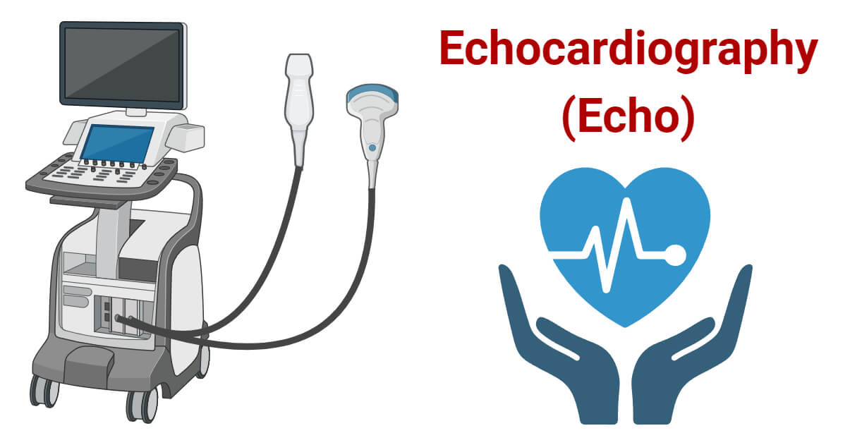
An echocardiography system, often called an Echo machine or Echocardiograph, is a non-invasive medical imaging device that uses high-frequency sound waves (ultrasound) to produce detailed, real-time images and videos of the heart. Its primary function is to assess the heart’s structure, function, and hemodynamics (blood flow) without the need for surgery or radiation.
Think of it as a specialized, highly sophisticated “ultrasound for the heart.” It allows clinicians to see the heart’s chambers, valves, walls, and the major blood vessels connected to it, evaluating how well it pumps blood and identifying a wide range of cardiac abnormalities.
How it Works
The system operates on the principle of ultrasound. Here’s a simplified breakdown:
- Transmission: A handheld probe called a transducer is placed on the patient’s chest. It emits high-frequency sound waves (inaudible to humans) that travel into the body.
- Reflection: These sound waves bounce off (echo) the various structures of the heart.
- Reception: The same transducer picks up the returning echoes.
- Processing: The system’s computer processes the time it takes for the echoes to return and their intensity. This data is used to generate:
- 2D Images: Cross-sectional, moving “slices” of the heart.
- Doppler Signals: Color-coded maps and spectral tracings that visualize the speed and direction of blood flow, revealing leaks, obstructions, and pressure gradients.
- Display: The images and data are displayed in real-time on the system’s monitor for immediate interpretation.
Key Components
- Transducer/Probe: The most critical component. Different probes (e.g., Phased Array, Sector) with varying frequencies are used for specific applications (transthoracic, transesophageal).
- System Console/Base Unit: The central computer housing the processing hardware and software. It controls all functions.
- Display Monitor: A high-resolution screen for viewing images and measurements.
- User Interface: Includes a keyboard, trackball/touchpad, and often a touchscreen for controlling the system and annotating images.
- Software Suite: The “brain” of the system. It includes:
- Image Acquisition Software: For capturing and optimizing images.
- Measurement & Analysis Packages: Quantitative tools for calculating ejection fraction, valve areas, strain, etc.
- Reporting Software: To generate structured patient reports.
- Data Management & Storage: Internal hard drives and connectivity (e.g., DICOM, HL7) to archive images to hospital servers (PACS) and integrate with Electronic Health Records (EHR).
- Patient Monitor (Optional on some systems): For simultaneous display of ECG, blood pressure, and oxygen saturation.
2. Uses
Clinical Applications
Echocardiography is the most widely used cardiac imaging modality.
- Diagnosis: Heart failure, cardiomyopathies, congenital heart disease, pericardial disease, cardiac masses (e.g., tumors, clots).
- Valvular Assessment: Stenosis (narrowing), regurgitation (leakage), prolapse, and endocarditis (infection).
- Quantification of Function: Measuring Ejection Fraction (EF) – the percentage of blood pumped out with each heartbeat – is a key application.
- Hemodynamic Assessment: Estimating pressures within the heart chambers and lungs (e.g., pulmonary artery pressure).
- Guidance: For procedures like pericardiocentesis (draining fluid) and during certain cardiac surgeries (Transesophageal Echo – TEE).
- Stress Echocardiography: Assessing heart function under physical or pharmacological stress to diagnose coronary artery disease.
Who Uses It
- Cardiologists & Pediatric Cardiologists: Primary interpreters and advanced users.
- Cardiac Sonographers/Echocardiographers: Specially trained technicians who perform the scans.
- Cardiothoracic Surgeons & Anesthesiologists: Often use TEE in the operating room.
- Intensivists & Emergency Physicians: Use point-of-care echocardiography (POCUS) for rapid assessment in critical settings.
Departments/Settings
- Cardiology Departments/Non-Invasive Labs
- Hospitals: Emergency Rooms (ER), Intensive Care Units (ICU), Operating Rooms (OR), and general wards.
- Outpatient Clinics & Diagnostic Centers
- Ambulatory Surgical Centers
- Mobile/Echo-Cart Systems for bedside use.
3. Technical Specs
Typical Specifications
- Imaging Modes: 2D, M-Mode, Color Doppler, Pulsed-Wave (PW) Doppler, Continuous-Wave (CW) Doppler, Tissue Doppler Imaging (TDI).
- Advanced Capabilities: 3D/4D Imaging, Speckle Tracking Echocardiography (Strain Imaging), Contrast Imaging.
- Probe Frequency Range: Typically 1-15 MHz. Lower frequencies (1-5 MHz) for deeper penetration (adults), higher frequencies (5-15 MHz) for better resolution of superficial structures (pediatrics, TEE).
- Display: High-definition (often 19″ or larger) LED/LCD monitors.
- Portability: Systems range from large, high-end carts to laptop-sized portable/handheld units.
Variants & Sizes
- High-End/Premium Cart-Based Systems: Top-tier image quality, all advanced features. Used in main echo labs.
- Mid-Range/Performance Cart-Based Systems: Excellent image quality for most clinical needs, more compact.
- Portable Systems: Lightweight, wheeled systems for bedside and point-of-care use.
- Handheld Ultrasound Devices (HUDs): Pocket-sized, transducer-connected to a smartphone/tablet. Used for rapid triage and basic assessment.
Materials & Features
- Construction: Durable, medical-grade plastics and metals designed for frequent cleaning.
- Transducers: Crystal arrays housed in ergonomic, sealed casings.
- Key Features: Wireless/Wi-Fi connectivity, AI-powered automation (auto-measurements, image optimization), cloud-based data sharing, enhanced workflow software.
Notable Models (Examples)
- GE HealthCare: Vivid E95 (Premium), Vivid E90, Vivid iq (Portable).
- Philips: EPIQ CVx, Affiniti CVx, Lumify (HUD).
- Siemens Healthineers: Acuson Sequoia, Acuson Juniper.
- Canon Medical Systems: Aplio i-series, Viamo.
- Fujifilm: iSpeed, iLog.
4. Benefits & Risks
Advantages
- Non-invasive & Painless: No needles, catheters, or incisions.
- No Ionizing Radiation: Safe for repeated use, including in pregnant women and children.
- Real-Time & Dynamic: Shows the heart beating and blood flowing.
- Highly Versatile & Comprehensive: Provides anatomical, functional, and hemodynamic data.
- Cost-Effective: Compared to CT or MRI.
- Portable: Can be brought to the patient’s bedside.
Limitations
- Acoustic Windows: Image quality can be poor in patients with obesity, lung disease, or certain chest wall deformities.
- Operator Dependency: Quality of exam heavily relies on the skill of the sonographer.
- Limited Field of View: Primarily images the heart; not a whole-body tool.
Safety Concerns & Warnings
- Thermal & Mechanical Indices: The system monitors and displays safety indices. Operators must follow the ALARA principle (As Low As Reasonably Achievable) to minimize energy exposure.
- Transesophageal Echo (TEE): Carries risks of sedation, esophageal injury, and aspiration.
- Infection Control: Probes must be cleaned and disinfected/sterilized appropriately between patients to prevent cross-contamination.
Contraindications
- Transthoracic Echo (TTE): Virtually none. It is a very low-risk procedure.
- Transesophageal Echo (TEE): Absolute contraindications include esophageal obstruction, perforation, or severe bleeding disorder. Relative contraindications include cervical spine instability, recent gastric surgery, or severe respiratory compromise.
5. Regulation
Echocardiography systems are regulated as medical devices globally.
- FDA Class: Typically Class II (moderate to high risk). Some advanced software features or novel applications may be classified differently.
- EU MDR Class: Usually Class IIa or IIb.
- CDSCO Category (India): Category C (moderate-high risk device).
- PMDA (Japan): Regulated as “Specified Controlled Medical Devices” (Class II, III, or IV depending on features).
- ISO/IEC Standards:
- ISO 13485: Quality Management Systems for Medical Devices.
- IEC 60601-1: General safety requirements for medical electrical equipment.
- IEC 60601-2-37: Particular safety standards for ultrasound diagnostic equipment.
6. Maintenance
Cleaning & Sterilization
- System Console & Monitor: Wipe daily and after each use with a mild, hospital-grade disinfectant. Avoid excess moisture.
- Transducer (TTE): Wipe probe and cable with a disinfectant wipe (e.g., CaviWipes) approved for ultrasound probes after every patient. Low-level disinfection is standard for intact skin.
- TEE Probe: Requires high-level disinfection or sterilization (e.g., using an automated endoscope reprocessor – AER) after each use, as it contacts mucous membranes.
Reprocessing
Strict adherence to manufacturer’s Instructions for Use (IFU) for probe cleaning and disinfection is mandatory to prevent damage and ensure efficacy.
Calibration
- Annual/Preventive Maintenance (PM): Performed by certified service engineers. Includes checking acoustic output, Doppler accuracy, and system performance against phantoms.
- User Calibration: Limited; mainly involves running internal software diagnostics.
Storage
- Store in a clean, dry, temperature-controlled environment.
- Transducers should be hung or stored in a way that prevents cable damage and probe-head contact with surfaces.
7. Procurement Guide
How to Select the Device
Consider: Clinical Needs (echo lab vs. ICU vs. clinic), Budget, User Skill Level, and Growth Plans.
Quality Factors
- Image Quality: The paramount factor. Assess clarity, resolution, and Doppler sensitivity.
- Workflow Efficiency: Intuitive interface, automated tools, and streamlined reporting.
- Durability & Service: Manufacturer’s reliability and quality of local service support.
- Upgradability: Can software/transducers be added later?
Certifications
Look for CE Marking (EU), FDA 510(k) Clearance (US), and local regulatory approvals (e.g., CDSCO, PMDA).
Compatibility
Must support DICOM and HL7 standards for seamless integration with PACS, EHR, and reporting systems.
Typical Pricing Range
- High-End Systems: $150,000 – $300,000+
- Mid-Range Systems: $70,000 – $150,000
- Portable Systems: $30,000 – $70,000
- Handheld Devices: $5,000 – $15,000 (probe + software)
Prices vary significantly based on configuration and region.
8. Top 10 Manufacturers (Worldwide)
- GE HealthCare (USA) – A global leader with the Vivid series. Known for premium image quality and advanced 4D/Strain technology.
- Philips (Netherlands) – Strong in cardiology with the EPIQ and Affiniti platforms, offering excellent workflow solutions.
- Siemens Healthineers (Germany) – Renowned for its Acuson Sequoia and Juniper systems, praised for exceptional 2D image clarity.
- Canon Medical Systems (Japan) – Formerly Toshiba. The Aplio i-series is highly regarded for its imaging precision and innovative applications.
- Fujifilm (Japan) – Gaining market share with its iSpeed and iLog systems, focusing on compact design and AI.
- Esaote (Italy) – A specialist in ultrasound, with a strong focus on cardiovascular and portable systems.
- Mindray (China) – A rapidly growing global player offering high-value, full-featured systems like the Resona series.
- Samsung Medison (South Korea) – Known for its HM70A and RS series, providing robust performance at competitive prices.
- Chison Medical Technologies (China) – A significant exporter of cost-effective ultrasound systems, including echocardiography.
- Butterfly Network (USA) – A disruptor with a single-probe, whole-body handheld ultrasound system (Butterfly iQ+) that includes cardiac capabilities.
9. Top 10 Exporting Countries (Latest Year – Based on Trade Data Trends)
- United States – Home to GE, Philips (HQ for healthcare), and Butterfly. The largest exporter of high-end systems.
- Netherlands – Major export hub for Philips’ global distribution.
- Germany – Home base for Siemens Healthineers, exporting premium technology worldwide.
- Japan – Canon and Fujifilm are key exporters of high-quality imaging systems across Asia and the West.
- China – A massive exporter of mid-range and economy systems from Mindray, Chison, and others.
- South Korea – Samsung Medison drives significant exports, particularly to emerging markets.
- Italy – Esaote is a notable exporter within Europe and to select global markets.
- Mexico – A growing manufacturing and export hub for several major brands serving the Americas.
- Singapore – A key regional distribution and logistics hub for medical devices in Asia-Pacific.
- United Kingdom – Exports specialized systems and software, despite being a large importer.
10. Market Trends
- Current Global Trends: Shift towards portability and point-of-care ultrasound. Increased integration of Artificial Intelligence (AI) for auto-measurements, guidance, and diagnosis. Growing demand in emerging markets.
- New Technologies: AI-driven workflow automation, 3D printing integration for procedural planning, cloud-based image sharing and collaboration, and ultrasound elastography for tissue characterization.
- Demand Drivers: Aging global population (increasing heart disease prevalence), rising minimally invasive cardiac procedures requiring imaging guidance, and the need for cost-effective diagnostics.
- Future Insights: Handheld devices will become more capable, blurring lines with cart-based systems. AI will move from an assistive tool to a more central role in quantitative analysis and quality control. Tele-echocardiography will expand access to expert care in remote areas.
11. Training
Required Competency
- Cardiac Sonographers: Require formal education (associate’s or bachelor’s degree) and certification (e.g., RDCS from ARDMS in the US).
- Physicians: Require structured fellowship training and credentialing in echocardiography (e.g., ASCeXAM by NBE).
- Point-of-Care Users: Require focused training courses (e.g., FATE, POCUS) to answer specific clinical questions.
Common User Errors
- Poor Image Optimization: Using default settings instead of adjusting gain, depth, and focus.
- Incorrect Doppler Alignment: Not aligning the Doppler beam parallel to blood flow, leading to inaccurate velocity measurements.
- Incomplete Exam: Failing to acquire all standard views and measurements.
- Probe Pressure: Applying excessive pressure, causing patient discomfort and suboptimal images.
Best-Practice Tips
- Start with a System Check: Ensure your probe and preset are correct for the exam.
- Follow a Protocol: Acquire images in a consistent, standardized sequence.
- Optimize, Then Freeze: Always optimize the 2D image before freezing to take measurements.
- Label Everything Clearly: Proper annotation is critical for interpretation and follow-up.
12. FAQs
- What’s the difference between an ECG and an Echo?
- ECG (Electrocardiogram) records the heart’s electrical activity (rhythm). Echo shows the heart’s physical structure, movement, and blood flow (pump function).
- Is the echocardiogram test painful?
- No. You may feel slight pressure from the transducer and coolness from the gel, but it is not painful.
- How long does an echocardiogram take?
- A standard Transthoracic Echo (TTE) typically takes 30 to 60 minutes.
- Can I eat or drink before an echo?
- For a standard TTE, yes. For a Transesophageal Echo (TEE), you will need to fast for several hours before the procedure.
- What does “ejection fraction” mean?
- It’s the percentage of blood pumped out of the left ventricle with each heartbeat. A normal EF is usually between 55% and 70%.
- Are there any side effects from the ultrasound waves?
- Diagnostic ultrasound is considered extremely safe with no known harmful side effects at the energies used.
- Why do I need a TEE if I already had a regular echo?
- The TEE probe gets much closer to the heart, providing clearer images of structures like heart valves, clots, or defects that may be hard to see from the chest wall.
- Can echo detect all heart problems?
- It is excellent for many issues but not all. For example, it may not visualize the coronary arteries directly (a CT angiogram does that) or very small electrical abnormalities.
- How often should an echo be repeated?
- It depends entirely on your specific heart condition. Your cardiologist will determine the appropriate follow-up interval.
- What is “Strain Imaging”?
- It’s an advanced echocardiography technique that measures the deformation (squeezing) of the heart muscle, often detecting subtle dysfunction before the ejection fraction drops.
13. Conclusion
The echocardiography system is a cornerstone of modern cardiovascular medicine. Its unique combination of safety, real-time imaging, comprehensive data, and versatility makes it an indispensable tool for diagnosing, managing, and monitoring heart disease across all clinical settings. From high-end lab systems to pocket-sized handheld devices, ongoing technological advancements—particularly in portability, AI, and connectivity—continue to expand its capabilities and accessibility. Successful implementation relies not only on choosing the right technology but also on investing in proper training, maintenance, and adherence to best practices, ensuring optimal patient care and outcomes.
14. References
- American Society of Echocardiography (ASE). Guidelines and Standards. https://www.asecho.org/
- U.S. Food and Drug Administration (FDA). Information for Ultrasound Imaging. https://www.fda.gov/radiation-emitting-products/medical-imaging/ultrasound-imaging
- Otto, C. M. (2019). Textbook of Clinical Echocardiography (7th ed.). Elsevier.
- World Health Organization (WHO). Medical Devices. https://www.who.int/health-topics/medical-devices
- Global Market Insights. Echocardiography Devices Market Report. (2023-2032).
- European Society of Cardiology (ESC). Cardiovascular Imaging. https://www.escardio.org/
- Manufacturer Websites: GE HealthCare, Philips, Siemens Healthineers, Canon Medical Systems.