
Introduction to Arthodesis Ankle Joint Fusion
Ankle arthrodesis, also known as ankle joint fusion, is a surgical procedure whereby the articular surfaces of the ankle joint (typically between the tibia and talus, sometimes incorporating parts of the fibula) are permanently fused together. Over time, the bones heal into one continuous bony bridge, eliminating motion at that joint. The main aim is to relieve pain caused by advanced joint degeneration or deformity, while maintaining a stable, plantigrade (flat-to-the-ground) foot for walking.
Though the concept of fusing joints is old, ankle arthrodesis remains a mainstay—or “gold standard”—for many patients with end-stage ankle arthritis, particularly when other treatments fail or are unsuitable. Even with the rise of total ankle replacement (arthroplasty), fusion continues to be more commonly performed in many settings due to its durability, fewer late revisions, and predictable outcomes.
That said, ankle fusion is not without significant trade-offs. The procedure sacrifices motion, places increased load on adjacent joints, and carries the risk of nonunion or malalignment. Hence, patient selection, surgical technique, and postoperative care are all crucial to success.
In this article, we explore the causes and indications that lead to fusion, the signs and diagnosis, detailed treatment approaches (including open, arthroscopic, hybrid), rehabilitation, complications, and long-term outcomes and patient perspectives.
Causes, Indications Risk Factors
Because arthrodesis is a treatment, not a disease, in this section “causes” really refers to the joint pathologies and patient factors that lead to the indication for fusion.
Indications: When Is Ankle Fusion Considered?
Ankle arthrodesis is typically indicated in end-stage ankle joint pathology, especially when pain and disability cannot be controlled by nonoperative means. Some of the common underlying causes include:
-
End-stage osteoarthritis (OA) of the ankle
-
Primary idiopathic OA is relatively rare in the ankle; most are secondary.
-
In advanced disease, cartilage loss, joint narrowing, osteophyte formation, and bone remodeling make joint-preserving options inadequate.
-
-
Post-traumatic arthritis
-
The most frequent cause of disabling ankle arthritis.
-
Prior fractures (e.g., tibial plafond, talus, malleoli), malunions, articular surface damage accelerate degeneration.
-
-
Failed prior ankle surgery / salvage of failed total ankle replacement (TAR or TAA)
-
If prior internal fixation, osteotomies, or arthroplasty fails, fusion may be the salvage solution.
-
-
Avascular necrosis (AVN) / collapse of talus or tibial plafond
-
Bone death leads to collapse, joint incongruity, fragmentation, and degeneration.
-
-
Neuropathic (Charcot) arthropathy
-
In diabetics or neuropathy patients, joint destruction may be too advanced for reconstruction.
-
-
Inflammatory or erosive arthropathies
-
Rare, but in some severe cases, the joint is too damaged for conservative measures.
-
-
Severe deformity or instability
-
When reconstruction is impossible due to malalignment, soft tissue deficiency, or bone loss.
-
Thus, fusion is often the “last resort” after many conservative and reconstructive options have been exhausted.
Risk Factors That Make Fusion Challenging (“Complex Fusion”)
Some patients are considered “complex fusion” candidates due to higher risk of complications (nonunion, infection, wound problems). A review of complex ankle arthrodesis outlines systemic and local risk factors.
Systemic risk factors include:
-
Smoking (strongly associated with higher rates of nonunion)
-
Advanced age
-
Diabetes (especially poorly controlled)
-
Peripheral vascular disease or poor circulation
-
Immune suppression or systemic inflammatory disease
-
Obesity, malnutrition, alcohol abuse
-
Poor patient compliance / nonadherence to weight-bearing restrictions
Local and anatomical risk factors include:
-
Poor soft-tissue envelope (thin skin, prior scars, prior surgeries, poor vascular supply)
-
Previous infection in the ankle or foot
-
Significant bone defects or loss
-
Malalignment, cavus or flatfoot deformities
-
Coexisting hindfoot pathology (e.g. subtalar arthritis)
-
Bone quality issues (osteoporosis)
In such challenging cases, the surgeon may use adjuncts (biological stimulators, staged operations, external fixators) to enhance success.
Symptoms and Signs Arthodesis Ankle Joint Fusion
When a patient is progressing toward or is a candidate for ankle arthrodesis, their symptoms often represent end-stage joint disease, not of the fusion itself.
Typical Symptoms
-
Persistent, often daily pain in the ankle region, worsened by weight-bearing (walking, stair climbing)
-
Pain in the front, medial or lateral ankle joint line
-
Swelling or intermittent joint effusions
-
Stiffness, restricted ankle motion (especially dorsiflexion/plantarflexion)
-
Grinding, crepitus, or grinding sensation with attempted motion
-
Instability, episodes of “giving way” if joint is lax or damaged
-
Difficulty walking on uneven ground or inclines
-
Reduced walking distance, limp, reliance on assistive devices
-
Night pain or rest pain in severe cases
Physical Examination Findings
-
Tenderness over the ankle joint line
-
Reduced or absent ankle dorsiflexion/plantarflexion
-
Deformity: varus, valgus, or rotational malalignment
-
Perimalleolar muscle atrophy
-
Altered gait pattern
-
Examination of adjacent joints (subtalar, midfoot) often reveals compensatory motion or early degeneration
-
Assessment of vascular status, skin integrity, prior scar lines
Given that fusion is a solution to advanced disease, the presentation tends to reflect long-standing, progressive degradation. The decision to fuse is made when symptom burden outweighs the functional loss from fusion.
Diagnostic Evaluation Preoperative Planning
A successful ankle fusion hinges on a thorough and multifaceted preoperative evaluation. This includes imaging, biomechanical planning, functional assessment, and medical optimization.
Imaging Modalities & Their Roles
-
Weight-Bearing Plain Radiographs
-
Essential first step: AP, lateral, mortise views under load.
-
Evaluate joint space narrowing, osteophytes, subchondral sclerosis, cysts, bone alignment.
-
Check for involvement of adjacent joints (subtalar, talonavicular, midfoot).
-
-
Computed Tomography (CT) Scan
-
Crucial for defining bony architecture, assessing defects, cysts, bone irregularities.
-
Helps in mapping fusion surfaces and planning grafts or bone cuts.
-
-
Magnetic Resonance Imaging (MRI)
-
Useful for soft tissue, cartilage remnants, ligament status, bone marrow edema, occult pathology.
-
In cases of suspected osteonecrosis, MRI helps assess viability.
-
-
Bone Scan / SPECT / Nuclear Imaging
-
May be helpful in evaluating activity or viability in bone, or to rule out infection.
-
-
Vascular Studies / Angiography
-
In borderline circulation cases, an angiogram may be warranted to assess blood flow.
-
-
Weight-bearing CT or advanced imaging
-
Some centers use weight-bearing CT for assessment of alignment and thresholds under load.
-
Biomechanical & Functional Assessment
-
Foot and ankle alignment — the goal in fusion is to achieve near-neutral alignment (slightly valgus, neutral dorsiflexion, slight external rotation) to optimize gait and reduce stress on adjacent joints.
-
Forefoot balance — forefoot varus/valgus or midfoot deformities need correction or compensation to avoid undue stresses post-fusion.
-
Adjacency joint health — checking for preexisting arthritis in subtalar, midfoot, talonavicular, calcaneocuboid joints
-
Leg-length discrepancy — fusions can introduce or amplify length differences; anticipate and plan accordingly
-
Gait and load distribution — preoperative gait analysis helps in understanding compensations and predicting outcomes
-
Bone quality evaluation — DEXA scans if there is suspicion of osteoporosis
-
Soft tissue envelope — assessing scars, skin quality, previous incisions, vascularity, and infection risk
-
Medical optimization — ensure comorbidities (diabetes, vascular disease, anemia, nutrition) are under control
-
Patient counseling and expectation management — discuss trade-offs, recovery timeline, functional limitations, possible need for revision
All this planning informs the surgeon's choice of approach, fixation, graft need, adjuncts, and postoperative protocols.
Treatment Options & Surgical Techniques
This is the core section. It describes the array of surgical options (and their pros/cons), decision-making, and technical pearls.
Nonoperative / Conservative Approaches (Before Fusion Decision)
Before a surgeon recommends fusion, almost all patients will have undergone a period of nonoperative management, which may include:
-
Rest, activity modification
-
Analgesics / NSAIDs
-
Bracing, orthoses (ankle-foot orthoses or supportive boots)
-
Physical therapy (strengthening, flexibility, gait training)
-
Intra-articular injections (e.g. corticosteroids, viscosupplementation)
-
Offloading via assistive devices (canes, crutches)
-
Shock-absorbing footwear or customized insoles
If these fail to yield acceptable pain relief or functional improvement, fusion becomes a viable option.
Surgical Fusion Approaches
The main dichotomy is open fusion vs arthroscopic (minimally invasive) fusion, with hybrid approaches or adjunct techniques for more complex cases. There is no clear consensus on a “best” method; choice depends on patient anatomy, deformity, surgeon experience, and bone/soft-tissue conditions.
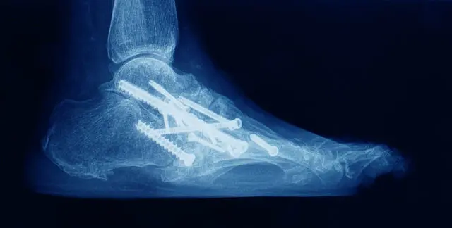
Open Ankle Arthrodesis
When to use it
-
Moderate to severe deformity (coronal/ sagittal)
-
Significant bone defects or need for grafting
-
Poor bone exposure via arthroscopic means
-
Complex revisions or salvage fusion
Common approaches
-
Anterior approach
-
Lateral (transfibular) approach
-
Posterior approach
-
Combined medial/lateral approaches
-
Sometimes dual incisions for severe deformities
Fixation options
-
Multiple large-diameter screws (often 2-3)
-
Plate + screws constructs (especially anterior plating)
-
Hybrid screw + plate designs
-
Supplemental external fixation in challenging cases
-
Bone grafting (autograft / allograft / bone substitutes) where defects exist
Technical considerations
-
Adequate preparation of joint surfaces (remove cartilage, sclerotic bone)
-
Compression across fusion interface
-
Correct alignment (valgus, neutral dorsiflexion, slight external rotation)
-
Maintenance of limb length and leg axis
-
Rigid fixation to reduce micromotion
-
Consider biologic augmentation in risk cases
Advantages
-
Better ability to correct deformities
-
Easier visualization and manipulation of bone/grafts
-
Reliable hardware application
Disadvantages / Risks
-
More soft tissue disruption
-
Potential for greater blood loss, wound healing issues
-
Longer hospital stay
In a systematic review, open approaches showed varied union and complication rates depending on approach and fixation.
Arthroscopic (Minimally Invasive) Ankle Arthrodesis
When considered
-
Mild-to-moderate deformity (e.g. ≤15° varus/valgus)
-
Intact bone stock without large defects
-
Favorable soft-tissue conditions
-
In experienced hands
Technique overview
-
Small portals with arthroscope + instrumentation
-
Cartilage removal and bone preparation under arthroscopic vision
-
Percutaneous screw fixation
-
Minimal soft-tissue disruption
Advantages
-
Less soft-tissue trauma, faster recovery
-
Less hospital stay and pain
-
Potential for faster bone healing initiation
-
Some studies show lower infection or wound complication rates.
Limitations / Risks
-
Limited ability to correct major deformities
-
Difficulty with significant bone defects, grafting, or irregular anatomy
-
Requires surgeon skill and familiarity
-
In some cases, adjacent joint stress may accelerate wear regardless of approach.
Recent evidence (JBJS) suggests that arthroscopic fusion has similar revision rates but lower infection rates compared to open fusion, though patient selection is important.
Hybrid / Combined Approaches & Alternatives
-
Open + arthroscopic hybrid: in selected cases, combining exposure and minimal invasiveness
-
Tibiotalocalcaneal (TTC) fusion / intramedullary nail: when subtalar joint also requires fusion or for severe deformity/instability
-
External fixation-assisted fusion: in cases of poor soft tissue or infection risk, staged use of external frames may be employed
-
Staged fusion: in cases with infection or bone loss, the surgeon may stage debridement, bone grafting, and final fusion
-
Bone stimulators / biophysical adjuncts: pulsed electromagnetic fields (PEMF), low-intensity ultrasound, direct-current stimulators to promote union, especially in “high-risk” fusion cases.
Evidence on Approaches & Outcomes
-
A meta-analysis comparing open versus arthroscopic fusion suggests arthroscopic has advantages of reduced soft-tissue trauma and hospital stay in selected cases.
-
Some newer comparative studies show little difference in long-term functional outcomes, but lower complication (infection) rates for arthroscopic fusion after adjusting baseline characteristics.
-
A recent retrospective cohort (2025) comparing anterior vs lateral approaches found no significant difference in union rate or complications, but stresses that surgical precision and technique (bone contact, alignment, fixation) are critical for success.
-
In anterior plating techniques, weight-bearing protocols did not significantly alter fusion rates.
-
Long-term outcomes: in one study of arthrodesis (43 women + 45 men), the overall complication rate was 11.4%, but 75% of patients reported full satisfaction.
It is important to note that ankle arthrodesis remains the predominant surgical option in many centers despite growing interest in ankle arthroplasty, largely because of its durability and more predictable long-term outcomes in many patient groups.
Postoperative Protocols & Rehabilitation
Proper postoperative care is as important as surgical technique. A generic staged protocol is as follows (to be adapted to surgeon's preference and patient factors):
-
Immobilization & Protection
-
Immediately postoperative: cast, boot, or external support
-
Strict non-weight bearing (duration depends on surgeon and fusion stability)
-
-
Wound & Infection Management
-
Antibiotic prophylaxis
-
Wound inspections, drainage monitoring
-
Vigilance for signs of infection
-
-
Edema Control & DVT Prevention
-
Elevation, cryotherapy, compression
-
Pharmacologic DVT prophylaxis as per protocol
-
-
Progressive Weight-Bearing
-
Partial weight-bearing (e.g. 6-8 weeks) if radiographic evidence of fusion onset
-
Gradual progression to full weight-bearing over months
-
-
Physical Therapy & Rehabilitation
-
Early: maintain mobility of adjacent joints (subtalar, midfoot, toes)
-
Later: strengthen muscles (calf, peroneals, intrinsic foot), balance and proprioception
-
Gait training and compensatory strategies
-
Use of orthotics or shoe modifications as needed
-
-
Serial Radiographic Surveillance
-
Radiographs (AP/lateral) at 6, 12, 24 weeks (or per protocol) to confirm union and detect malalignment or hardware problems
-
CT may be used if union is uncertain
-
-
Lifestyle & Behavioral Guidance
-
Avoid high-impact or torsional stress
-
Appropriate footwear (shock absorption, stability)
-
Weight management
-
Ongoing monitoring of adjacent joints and any symptoms
-
-
Use of Adjuncts if Fusion Risk Is High
-
Bone stimulators (ultrasound, electromagnetic) when nonunion risk is high
-
Supplementation or nutrition optimization
-
Adherence to rehabilitation, weight-bearing restrictions, and follow-up is critical. Noncompliance is a known risk factor for nonunion or hardware failure.
Prevention and Management Strategies (Optimizing Outcomes)
While you cannot completely prevent the need for fusion in someone with advanced arthritis, many strategies help reduce complications and optimize fusion success.
Preoperative Measures / Preventive Optimization
-
Smoking cessation, ideally several weeks to months before surgery
-
Glycemic control and optimization of comorbidities (diabetes, anemia, vascular disease)
-
Nutrition optimization, including protein status, vitamin D, bone health
-
Vascular evaluation in marginal perfusion cases
-
Soft tissue planning: ensure that scar lines, skin flaps, prior incisions are incorporated appropriately
-
Stabilizing adjacent deformities (forefoot, hindfoot) ahead of or concurrently
-
Patient counseling: set realistic expectations, emphasize compliance
Intraoperative Techniques to Prevent Complications
-
Precise cartilage removal and decortication to healthy bleeding bone
-
Maximal compression and stable fixation
-
Rigid fixation (screws, plates) with minimal micro-motion
-
Addressing bone defects aggressively (grafts, structural support)
-
Judicious use of biologic adjuncts (bone morphogenetic proteins, bone graft substitutes) in high-risk cases
-
Minimize soft-tissue trauma
-
Meticulous hemostasis
-
Prevent overcorrection or malalignment
Postoperative Risk Mitigation
-
Strict adherence to weight-bearing protocols
-
Close monitoring of wound, early treatment of complications
-
Edema control and DVT prophylaxis
-
Early identification of union delays or hardware problems via imaging
-
Use of bone stimulators if delayed union suspected
-
Prompt revision when necessary
Preventing nonunion, infection, malalignment, or hardware failure is a multi-step, multidisciplinary process involving surgeon skill, patient selection, and postoperative care.
Potential Complications & How They Are Managed
Ankle fusion is a major surgery and carries a meaningful risk profile. Recognizing complications and planning to mitigate them is essential.
Common & Significant Complications
-
Nonunion / Delayed Union
-
Failure of bony fusion
-
Risk factors: smoking, poor vascularity, comorbidities, micromotion
-
Management: revision fusion, bone grafting, biologics, re-fixation
-
-
Infection / Wound Healing Problems
-
Superficial or deep surgical site infection
-
Delayed wound closure or dehiscence
-
Management: debridement, antibiotics, staged revision
-
-
Hardware Problems
-
Loosening, breakage, prominence, irritation
-
May require hardware removal or revision
-
-
Malalignment / Malposition / Varus-Valgus Deviation
-
Poor alignment leads to abnormal gait or pain
-
May require corrective osteotomy or revision
-
-
Adjacent Joint Arthritis / Degeneration
-
The loss of ankle motion transfers stress to subtalar, midfoot, talonavicular joints
-
Over time, this may lead to arthritis and symptoms
-
-
Stress Fractures in Adjacent Bones
-
Secondary to altered load transmission
-
-
Neurovascular Injury
-
Nerve damage, vascular compromise during surgery
-
-
Deep Vein Thrombosis (DVT) / Pulmonary Embolism (PE)
-
Standard prophylaxis needed
-
-
Persistent Pain / Partial Relief
-
Even when fused, some patients continue to have pain due to other factors
-
-
Reoperation / Revision Surgery
-
Nonunion, malalignment, hardware failure often lead to re-operation
-
-
Amputation (Rare, in extreme failure / infection)
-
Rare but catastrophic in salvage failure cases
-
In one clinical series, the overall complication rate was 11.4%, with delayed wound healing, infection, and nonunion being the most frequent.
A review of complex fusions notes that the rate of nonunion and wound problems is higher in “complex” patients (comorbidities, poor soft tissues, bone defects).
Thus, prompt recognition, imaging surveillance, and readiness to intervene are keys to optimal outcomes.
Living with the Condition of Arthodesis Ankle Joint Fusion
This section addresses what patients can expect long-term, how to manage life with a fused ankle, and strategies to optimize function and satisfaction.
Functional Outcomes & Patient Satisfaction
-
Many studies show that ankle arthrodesis reliably relieves pain and improves function. In the aforementioned series, 75% of patients reported full satisfaction.
-
The American Orthopaedic Foot & Ankle Society (AOFAS) scores often improve significantly post-fusion.
-
However, patients lose ankle dorsiflexion/plantarflexion; the motion must be compensated by adjacent joints (subtalar, midfoot).
-
Studies comparing open vs arthroscopic fusion show similar long-term outcomes, though arthroscopic fusion may lead to faster early improvement and fewer soft-tissue complications.
-
The incidence of adjacent joint arthritis increases with time and is a recognized long-term trade-off.
Long-Term Considerations: Adjacent Joint Degeneration & Salvage Options
-
Because ankle motion is lost, loading shifts to the subtalar joint, talonavicular joint, and midfoot joints. Over years, these joints may undergo degenerative changes, possibly leading to pain or secondary surgeries.
-
Longitudinal studies suggest that many patients remain satisfied for decades, but some will require additional treatment for adjacent joint problems.
-
If nonunion or failure occurs, revision fusion, bone grafting, or alternative salvage procedures are possible.
-
In limited settings, conversion to ankle arthroplasty (if anatomy and soft tissues permit) is occasionally reported, but is often technically complex and less commonly done.
Patient Adaptation & Quality of Life
Patients living with a fused ankle adapt in several ways:
-
Footwear modifications — shock-absorbing soles, rocker soles, cushioned insoles
-
Orthoses or custom inserts to distribute load
-
Avoidance of high-impact activities (running, jumping)
-
Strength, balance, flexibility training for adjacent joints
-
Monitoring for new pain in midfoot, hindfoot, knee, hip
-
Periodic evaluation for adjacent joint degeneration
Many patients return to daily life and many recreational activities (walking, cycling, swimming). While high-demand sports may be limited, patient satisfaction is generally good when expectations are aligned.
Prognosis & Survival
-
Union rates in modern series vary but commonly are in the 90%+ range under favorable conditions.
-
When fusion succeeds with good alignment and stable fixation, long-term durability is good
-
The primary long-term issues are adjacent joint degeneration and the possibility of revision operations
-
Patients with diabetes, older age, or comorbidities have higher complication rates, so their prognosis is more guarded
Top 10 Frequently Asked Questions about Ankle Arthrodesis (Ankle Joint Fusion)
1. What is Ankle Arthrodesis (Ankle Joint Fusion)?
Ankle arthrodesis, commonly called ankle joint fusion, is a surgical procedure that involves fusing the bones of the ankle joint together. The primary goal is to eliminate pain and stabilize the ankle, particularly in patients with advanced arthritis, severe deformities, or irreparable joint damage.
Unlike ankle replacement, which preserves joint motion, ankle arthrodesis results in loss of movement in the fused joint, but other joints in the foot and ankle help maintain functional mobility. This procedure is considered the gold standard for severe ankle arthritis when non-surgical treatments have failed.
2. Why is Ankle Arthrodesis Performed?
Ankle arthrodesis is typically recommended in the following scenarios:
-
Severe Osteoarthritis or Rheumatoid Arthritis: When the ankle joint cartilage is completely worn out.
-
Post-Traumatic Arthritis: Following fractures, ligament injuries, or previous failed surgeries.
-
Congenital or Acquired Deformities: To correct severe misalignment or instability.
-
Failed Ankle Replacement: When previous joint replacement surgery does not relieve pain.
-
Chronic Instability or Severe Pain: Which significantly affects walking, balance, or quality of life.
The procedure provides pain relief, joint stability, and improved walking ability, even though ankle motion is sacrificed.
3. What Does the Surgery Involve?
Ankle arthrodesis can be performed using several surgical techniques, including open fusion, arthroscopic fusion, and minimally invasive approaches. The general steps include:
-
Anesthesia: General or spinal anesthesia is administered.
-
Incision: Depending on technique, the surgeon makes an incision over the ankle joint.
-
Cartilage Removal: Damaged cartilage is removed from the joint surfaces.
-
Bone Alignment: The tibia and talus bones are positioned in a natural alignment.
-
Fixation: Screws, plates, or rods are used to hold the bones together.
-
Closure: The incision is closed with sutures, and a sterile dressing is applied.
The surgery typically takes 2-3 hours, depending on complexity.
4. What Are the Benefits of Ankle Arthrodesis?
The main benefits include:
-
Pain Relief: Significantly reduces or eliminates chronic ankle pain.
-
Joint Stability: Corrects deformities and stabilizes the ankle.
-
Improved Function: Patients can walk comfortably and perform daily activities.
-
High Success Rate: Long-term results are generally excellent, with most patients reporting satisfaction.
While ankle motion is lost, the procedure provides durable pain relief and improved quality of life for patients with end-stage ankle disease.
5. What Are the Risks and Complications?
As with any major surgery, ankle arthrodesis carries potential risks:
-
Nonunion or Delayed Healing: Occurs when the bones fail to fuse completely.
-
Infection: At the surgical site, sometimes requiring additional treatment.
-
Nerve or Blood Vessel Injury: Rare but possible.
-
Changes in Gait: Compensatory changes in walking may cause stress on other joints.
-
Hardware Problems: Screws or plates may loosen or irritate surrounding tissue.
-
Blood Clots (DVT): Deep vein thrombosis may occur post-operatively.
Choosing an experienced orthopedic surgeon and adhering to post-operative care minimizes complications.
6. What is the Recovery Process Like?
Recovery following ankle arthrodesis is gradual:
-
Hospital Stay: Usually 2-5 days, depending on complexity and patient health.
-
Immobilization: A cast or boot is applied to protect the fused joint, typically for 6-12 weeks.
-
Non-Weight Bearing: Patients use crutches or a walker during the initial healing period.
-
Physical Therapy: Begins gradually to strengthen surrounding muscles and improve mobility.
-
Return to Activities: Most patients can resume light daily activities within 3-4 months, while high-impact activities may remain restricted.
Full fusion and optimal outcomes may take 6-12 months.
7. Will I Lose All Movement in My Ankle?
Yes. After ankle arthrodesis, the fused joint will no longer move. However, other joints in the foot, such as the subtalar joint and midfoot joints, compensate for some motion, allowing patients to walk and perform normal activities. Most patients find that the pain relief outweighs the loss of motion, especially in severe arthritis.
8. Are There Alternatives to Ankle Arthrodesis?
Yes, alternatives may include:
-
Total Ankle Replacement (Ankle Arthroplasty): Preserves motion while relieving pain, suitable for selected patients.
-
Conservative Management: Physical therapy, bracing, pain medications, and injections may help in mild or moderate cases.
-
Osteotomy or Realignment Procedures: For younger patients with deformity but preserved cartilage.
The choice depends on patient age, activity level, bone quality, and severity of joint damage.
9. How Do I Know if I'm a Candidate for Ankle Arthrodesis?
Candidates typically have:
-
Severe ankle arthritis not relieved by conservative treatments.
-
Chronic pain affecting daily life and mobility.
-
Deformity or instability of the ankle joint.
-
Good overall health for surgery and rehabilitation.
A thorough consultation with an orthopedic surgeon, including X-rays or CT scans, helps determine suitability.
10. What is the Long-Term Outlook After Surgery?
-
Most patients experience long-lasting pain relief and improved function.
-
They are able to walk comfortably and resume daily activities.
-
Some patients may experience mild stress on adjacent joints, which can be managed with orthotics or physical therapy.
-
Proper follow-up and adherence to post-operative care ensure optimal outcomes and minimize complications.
While ankle arthrodesis eliminates motion at the fused joint, it remains a reliable, durable solution for patients with end-stage ankle disease.