
Introduction to Nephroureterectomy Bladder Cuff Excision
Nephroureterectomy with bladder cuff excision is a surgical procedure primarily used to treat cancers of the upper urinary tract, including the kidney and ureter, and to remove affected portions of the bladder. The surgery involves the removal of the kidney (nephrectomy), the ureter (the tube that connects the kidney to the bladder), and a portion of the bladder near the point where the ureter enters. This approach is most commonly used for urothelial carcinoma, a type of cancer that affects the lining of the urinary tract. It is also performed in cases of ureteral obstruction, severe damage, or recurrent infection when conservative treatments are ineffective.
The procedure is typically carried out under general anesthesia, and recovery can take several weeks. Following surgery, patients may need to manage urinary diversion and undergo regular follow-up care to monitor for any signs of cancer recurrence. Nephroureterectomy with bladder cuff excision provides a potentially curative option for patients with localized cancer, offering long-term survival benefits when carefully managed.
Causes and Risk Factors of Nephroureterectomy & Bladder Cuff Excision
Nephroureterectomy with bladder cuff excision is a surgical procedure primarily used for treating urinary tract cancers, including renal cell carcinoma, urothelial carcinoma, or transitional cell carcinoma that affects the kidney, ureter, and bladder. The procedure involves the removal of the kidney, ureter, and a portion of the bladder around the ureter's entrance (the bladder cuff). Understanding the causes of the need for this surgery and the associated risk factors is critical for managing and preventing complications.
1. Cancers Involving the Kidney and Ureter
-
Upper Tract Urothelial Carcinoma: This is the most common cancer type that necessitates nephroureterectomy. It often starts in the lining of the kidney or ureter and can be aggressive.
-
Renal Cell Carcinoma: This kidney cancer may extend to the ureter and surrounding tissues, requiring removal of the kidney, ureter, and part of the bladder.
-
Transitional Cell Carcinoma: A form of cancer affecting the urothelial lining of the kidney and ureter.
2. Chronic and Recurrent Infections
-
Chronic Pyelonephritis: Persistent kidney infections that may damage the kidney and ureter, leading to kidney failure and requiring nephrectomy.
-
Obstructive Uropathy: When an obstruction in the urinary tract leads to long-term kidney damage or infection, nephrectomy may be necessary to remove the damaged organ and restore proper function.
3. Trauma or Injury
-
Blunt Force Trauma: Significant physical injury to the kidney, such as from an accident or sports-related injury, may necessitate nephrectomy if the damage is severe.
-
Surgical Injury: During surgeries of the pelvic or abdominal regions, accidental injury to the kidney or ureter may require removal.
4. Congenital Abnormalities
-
Multicystic Dysplastic Kidney Disease: A condition in which cysts develop in the kidney, potentially leading to nephrectomy if the kidney is non-functional.
-
Ureteropelvic Junction Obstruction: A congenital condition that causes the kidney's urine flow to be obstructed, potentially requiring nephroureterectomy.
Symptoms and Signs of Nephroureterectomy & Bladder Cuff Excision
Nephroureterectomy and bladder cuff excision are surgical procedures primarily performed for the treatment of urinary tract cancers or conditions like upper tract urothelial carcinoma. The symptoms and signs associated with these conditions and the procedures can vary, but common symptoms include:
1. Pain and Discomfort
-
Flank Pain: Dull or sharp pain in the side or back, often felt below the ribs, is a classic sign of kidney problems or obstruction.
-
Pelvic Pain: If the ureter or bladder is involved, pain may radiate to the pelvic area.
2. Urinary Symptoms
-
Blood in Urine (Hematuria): A common sign of cancer or severe infection in the kidneys or ureter.
-
Frequent Urination: This can occur due to obstruction, infection, or cancer involving the urinary tract.
-
Painful Urination (Dysuria): A sign of infection, irritation, or obstruction in the ureter or bladder.
3. Systemic Symptoms
-
Weight Loss: Unexplained weight loss, especially in conjunction with pain, blood in the urine, or other urinary symptoms, may point to cancer.
-
Fatigue: Chronic kidney disease or kidney cancer often results in fatigue and general malaise.
4. Edema (Swelling)
-
Fluid Retention: Swelling in the legs, feet, or abdomen due to kidney dysfunction or urinary obstruction.
Diagnosis of Nephroureterectomy & Bladder Cuff Excision
Nephroureterectomy and bladder cuff excision are procedures commonly performed for certain types of urinary tract cancers, such as transitional cell carcinoma, which affect the kidney, ureter, or bladder. Accurate diagnosis is essential to determine the need for these surgeries and to ensure appropriate treatment planning.
1. Physical Examination
-
A thorough physical examination is performed to assess abdominal and flank tenderness, urinary symptoms, and hydration status.
2. Imaging Techniques
-
CT Scan (Computed Tomography): Essential for visualizing kidney and ureteral masses and determining the extent of cancer.
-
Ultrasound: A non-invasive imaging technique that helps evaluate kidney size, structure, and possible cysts or masses.
-
MRI (Magnetic Resonance Imaging): Provides high-resolution images of soft tissues, which is helpful in planning surgery.
3. Biopsy and Urine Tests
-
Urinary Cytology: A test to detect cancerous cells in the urine.
-
Biopsy: If cancer is suspected, a biopsy is performed to confirm the type of cancer and stage the disease.
4. Blood Tests
-
Renal Function Tests: These assess kidney function and identify signs of renal failure.
-
Tumor Markers: Blood tests to check for elevated levels of markers indicative of kidney or urothelial cancer.
Treatment Options of Nephroureterectomy & Bladder Cuff Excision
Nephroureterectomy and bladder cuff excision are surgical procedures typically performed to treat various urological conditions, including cancer or severe damage to the kidneys, ureters, or bladder. The treatment options for these conditions depend on the underlying cause, the patient's health, and the extent of the disease. Here's an overview of treatment options related to nephroureterectomy and bladder cuff excision:
1. Surgical Approaches
-
Radical Nephroureterectomy with Bladder Cuff Excision: The primary surgical option for upper tract urothelial carcinoma. This procedure involves removing the affected kidney, ureter, and a portion of the bladder.
-
Laparoscopic/Robotic-Assisted Surgery: Minimally invasive techniques have become increasingly popular, offering quicker recovery times and smaller incisions. Laparoscopic surgery is now commonly used for nephroureterectomy and bladder cuff excision.
-
Partial Nephrectomy: If the cancer is confined to a small portion of the kidney, only that portion may be removed, preserving the remaining healthy kidney tissue.
2. Post-Surgical Management
-
Urinary Diversion: After removal of the bladder cuff, urinary diversion techniques, including the use of a ureterostomy, may be required.
-
Pain Management: Post-operative pain is managed with analgesics, including non-steroidal anti-inflammatory drugs (NSAIDs) and opioid painkillers, if needed.
3. Adjuvant Therapies
-
Chemotherapy: Often used to treat urothelial carcinoma after surgery, particularly if cancer cells have spread to surrounding tissues.
-
Radiation Therapy: In some cases, radiation therapy may be used to target remaining cancer cells after surgical removal.
Prevention and Management of Nephroureterectomy & Bladder Cuff Excision
Nephroureterectomy (NUE) and bladder cuff excision are surgical procedures often performed to treat conditions such as upper tract urothelial carcinoma, severe obstructive uropathy, or other urinary tract cancers. The goal of these procedures is to remove the kidney, ureter, and sometimes a portion of the bladder cuff to prevent the spread of cancer or other diseases. Proper prevention and management before, during, and after the surgery are crucial for optimal recovery and outcomes.
1. Prevention
-
Lifestyle Modifications: Quitting smoking and reducing alcohol consumption can significantly reduce the risk of kidney and bladder cancers.
-
Regular Screenings: For those at high risk, such as individuals with a family history of cancer, regular screenings, including urine cytology and CT scans, are recommended.
2. Post-Operative Care
-
Hydration: Patients are advised to maintain optimal fluid intake post-surgery to aid kidney function and prevent complications such as kidney stones or infection.
-
Wound Care: Proper wound care is essential to prevent infection at the surgical site.
-
Physical Therapy: For patients who undergo laparoscopic surgery, early mobilization and physical therapy may be recommended to improve recovery.
Complications of Nephroureterectomy & Bladder Cuff Excision
Nephroureterectomy with bladder cuff excision is a significant surgical procedure typically performed for the treatment of upper urinary tract malignancies or in cases of severe urothelial cancers. While this procedure can be life-saving, it comes with potential complications that need to be carefully managed. Below are some of the major complications associated with nephroureterectomy and bladder cuff excision:
1. Infection
-
Urinary tract infections (UTIs) or surgical site infections can occur and may require antibiotics.
2. Bleeding
-
Excessive bleeding during or after the surgery can sometimes require blood transfusions or additional surgical interventions.
3. Kidney Dysfunction
-
After the removal of one kidney, the remaining kidney may have to work harder, potentially leading to renal insufficiency or acute kidney failure.
4. Urinary Incontinence
-
There is a possibility of incontinence after bladder cuff excision, especially if the bladder function is impaired.
Living with the Condition of Nephroureterectomy & Bladder Cuff Excision
Living with the condition after nephroureterectomy and bladder cuff excision involves adapting to significant changes in both physical and emotional health. This major surgery typically involves the removal of a kidney (or part of one) and the bladder cuff, which may lead to lifestyle adjustments, ongoing medical monitoring, and, in some cases, permanent changes to the body's normal functions. Here are the key aspects of life after this surgery:
1. Physical Adjustments
-
Kidney Function: With only one kidney remaining, it is important to manage hydration levels, limit salt intake, and maintain a healthy diet to support the remaining kidney's function.
-
Urinary Changes: Some patients may need a catheter for a temporary period following the surgery. Long-term urinary diversions may be required in certain cases.
2. Psychosocial Impact
-
The diagnosis of cancer and subsequent surgery can cause significant emotional distress. Psychological counseling and support groups are essential for helping patients cope with the emotional impact.
3. Long-Term Care
-
Survival and Follow-up: Regular follow-ups are required to monitor kidney function, detect potential recurrence of cancer, and manage any other long-term complications.
-
Physical Rehabilitation: Gradual rehabilitation helps patients regain strength and return to their daily activities.
Top 10 Frequently Asked Questions about Nephroureterectomy & Bladder Cuff Excision
1. What is nephroureterectomy with bladder cuff excision?
Nephroureterectomy with bladder cuff excision is a surgical procedure that involves the removal of the kidney, the entire ureter, and a small portion of the bladder where the ureter connects. This procedure is commonly performed to treat upper tract urothelial carcinoma (UTUC), a type of cancer affecting the renal pelvis and ureter. The goal is to eliminate the tumor and reduce the risk of recurrence.
2. Why is bladder cuff excision performed during nephroureterectomy?
Bladder cuff excision is performed to:
-
Remove potential cancerous tissue: Tumor cells may be present at the junction where the ureter enters the bladder.
-
Reduce recurrence risk: Removing this tissue decreases the likelihood of cancer returning in the bladder.
-
Improve oncological outcomes: Studies have shown that bladder cuff excision improves recurrence-free survival, particularly concerning bladder recurrence.
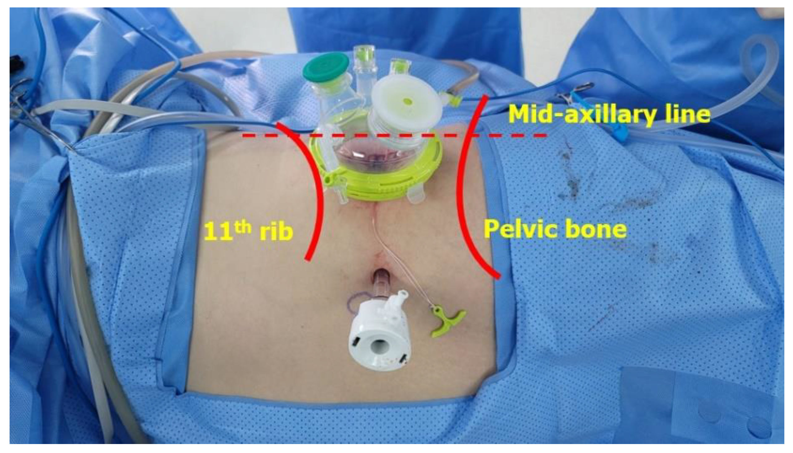
3. What are the surgical approaches to nephroureterectomy with bladder cuff excision?
The procedure can be performed using:
-
Open surgery: A single large incision is made to access and remove the kidney, ureter, and bladder cuff.
-
Laparoscopic surgery: Multiple small incisions are made, and the surgeon uses a camera and specialized instruments to perform the surgery.
-
Robotic-assisted surgery: A minimally invasive approach where the surgeon controls robotic instruments to perform the surgery with enhanced precision.
4. What are the benefits of robotic-assisted nephroureterectomy?
Robotic-assisted nephroureterectomy offers several advantages:
-
Minimally invasive: Smaller incisions lead to less pain and quicker recovery.
-
Enhanced precision: Robotic instruments provide greater dexterity and visualization.
-
Shorter hospital stay: Patients often experience a reduced length of hospitalization.
-
Faster recovery: Quicker return to normal activities compared to traditional open surgery.
5. What are the risks associated with the procedure?
As with any major surgery, risks include:
-
Infection: At the surgical site or internally.
-
Bleeding: During or after surgery.
-
Urinary complications: Such as leakage or obstruction.
-
Damage to surrounding organs: Including the bladder, blood vessels, or intestines.
-
Anesthesia risks: Reactions to anesthesia medications.
It's essential to discuss these risks with your healthcare provider to understand and mitigate them.
6. How long is the recovery period after surgery?
Recovery times can vary:
-
Hospital stay: Typically 2 to 5 days, depending on the surgical approach and individual recovery.
-
Return to normal activities: Most patients can resume normal activities within 4 to 6 weeks.
-
Follow-up care: Regular check-ups are necessary to monitor for any signs of recurrence or complications.
Your healthcare team will provide personalized guidance based on your specific situation.
7. What is the success rate of nephroureterectomy with bladder cuff excision?
The success rate varies based on factors like cancer stage and grade:
-
Overall survival: Studies report 5-year overall survival rates ranging from 60% to 80%.
-
Cancer-specific survival: Higher rates, often exceeding 80%, especially in early-stage cancers.
-
Recurrence-free survival: Bladder cuff excision has been shown to improve recurrence-free survival, particularly concerning bladder recurrence.
Early detection and appropriate surgical intervention are key to favorable outcomes.
8. Are there alternatives to this procedure?
Yes, alternatives include:
-
Endoscopic resection: For small, low-grade tumors confined to the renal pelvis or proximal ureter.
-
Segmental ureterectomy: Removal of the affected segment of the ureter, preserving kidney function.
-
Nephron-sparing surgery: Aims to remove the tumor while preserving as much kidney tissue as possible.
The choice of treatment depends on tumor characteristics, patient health, and surgeon expertise.
9. How should I prepare for the surgery?
Preparation includes:
-
Preoperative assessments: Blood tests, imaging studies, and possibly a cystoscopy.
-
Medication review: Adjusting or discontinuing certain medications as advised by your doctor.
-
Fasting: Avoiding food and drink for a specified period before surgery.
-
Anesthesia consultation: Discussing anesthesia options and any concerns.
Your surgical team will provide detailed instructions tailored to your situation.
10. When should I contact my healthcare provider post-surgery?
Contact your provider if you experience:
-
Signs of infection: Fever, redness, or discharge at the surgical site.
-
Urinary issues: Painful urination, blood in urine, or difficulty urinating.
-
Severe pain: Not controlled by prescribed medications.
-
Unusual swelling or bleeding: In the abdominal area or elsewhere.
Prompt attention to these symptoms can prevent complications and ensure a smooth recovery.